Quantcast
Channel: Imaging Technology News - Angiography
Viewing all 137 articles
Browse latest View live

WVU Medicine Installs First Alphenix 4D CT in the U.S.

$
0
0
WVU Medicine Installs First Alphenix 4D CT in the U.S.

July 24, 2019 — The West Virginia University (WVU) Heart and Vascular Institute is the first hospital in the country to acquire the Alphenix 4D CT from Canon Medical Systems USA Inc. The system offers an angiography configuration to expand capabilities in interventional procedures and help advance patient care in the community. The configuration pairs the Alphenix Sky + C-arm and Hybrid Catheterization Tilt/Cradle Table for interventional procedures with the Aquilion One/Genesis Edition computed tomography (CT) system, allowing clinicians to efficiently plan, treat and verify in a single clinical setting.

The WVU Heart and Vascular Institute uses the Alphenix 4D CT to perform interventional procedures for a wide range of applications, including advanced neurointerventional work, and to calculate diagnoses for heart and vascular diseases. In addition, the system is helping clinicians confidently identify and treat gastrointestinal (GI) bleeds, tumor lesions, percutaneous embolization and chest hematomas, among other complications.

“As we looked to upgrade our interventional radiology equipment, we first had to determine what is driving healthcare and found that a large focus is on heart disease, vascular disease and cancer,” said Luke Marone, M.D., co-director, chief of vascular interventional services, WVU Heart and Vascular Institute. “Already, the system has helped our clinicians think strategically about how they can leverage new technology to expand the horizons of care we offer.”

The Alphenix 4D CT is part of Canon Medical’s suite of Collaborative Imaging tools which puts integrated imaging intelligence at the center of a patient’s journey. The initiative fuses multiple diagnostic imaging modalities with clinical applications to deliver holistic, optimized patient information to healthcare providers at the point of care.

For more information: www.us.medical.canon

Wednesday, July 24, 2019 - 08:00

Jackson Memorial Hospital Holds Ribbon-cutting for New Cardiac Catheterization Labs

$
0
0
Jackson Memorial Hospital Holds Ribbon-cutting for New Cardiac Catheterization Labs

August 21, 2019 — Jackson Memorial Hospital in Miami celebrated the opening of two newly renovated cardiac catheterization suites during a ribbon-cutting ceremony on August 19, 2019. The suites feature the new Alphenix Core+ from Canon Medical Systems, which provides diverse imaging without compromising image quality or performance. Jackson Memorial is the first hospital in Florida offering this new technology.

“Almost six years ago, when Miami-Dade voters approved the bond, we promised a huge makeover for Jackson – new facilities, renovated patients units and new technology,” Carlos A. Migoya, president and CEO of Jackson Health System, said at the ribbon cutting. “We are so excited to launch these new catheterization labs, which offer the most sophisticated and advanced technology to our cardiology patients.”

Made with what Canon calls the world’s first high-definition detector for resolving fine details, the hybrid 12 x 12-inch panel on the Alphenix Core+ is combined with high-definition flat panel technology.

The Alphenix Hi-Def Detector technology helps clinicians see finer details during complex interventional procedures, such as stent positioning and stent apposition, wire and catheter navigation through the stent struts, and observation of coil deployment. The slim, off-center C-arm design on the Alphenix Core+ also allows steep angulations for optimized vessel profiling during cardiac interventions. In addition to offering high-quality imaging, the system has several components that allow for a wider range of procedures.

This state-of-the-art system, combined with the expertise of Jackson physicians, will provide patients with the highest quality experience and best outcomes, said Alexandre Ferreira, M.D., chief of cardiology and medical director of cardiology at Jackson Health System.

“We are so proud to unveil the new cardiac catheterization labs, which will be the primary treatment area for many diagnostics and interventional heart procedures,” said Ferreira. “With the use of the Alphenix Core+, patients will be able to receive a wider range of procedures without needing to be moved, while the care team access controls with a digital tablet. The rooms also offer 3-D imagery on live displays, and the lowest possible radiation doses.”

With this new technology, Jackson’s cardiology team can track in real time the estimated skin dose of X-ray exposure, which is displayed on the screen with designated colors ranging from lowest to the highest exposure. This is a vital patient safety feature.

Patients in need of procedures, such as angioplasty, stent implantation and heart catheterization, will be treated in these new cath labs. These minimally invasive tests and procedures can be performed instead of heart surgery to access heart and blood vessels.

Earlier this year, Jackson launched the Heart Surgery Institute, which combined a well-known group of cardiac surgeons with its existing cardiology team, providing the most comprehensive cardiac services in the region for patients of all ages.

For more information: www.jacksonhealth.org

Wednesday, August 21, 2019 - 13:30

Artis icono biplane

Artis icono floor

FDA Clears Artis Icono Family of Angiography Systems from Siemens Healthineers

$
0
0
The Artis icono biplane angiography system is designed for use in neurointerventions and interventional radiology

The Artis icono biplane angiography system is designed for use in neurointerventions and interventional radiology. Image courtesy of Siemens Healthineers.

The Artis icono floor angiography system is a flexible, multi-axis option for a wide range of disciplines, particularly vascular, interventional cardiology, surgical and interventional oncology

The Artis icono floor angiography system is a flexible, multi-axis option for a wide range of disciplines, particularly vascular, interventional cardiology, surgical and interventional oncology. Image courtesy of Siemens Healthineers.

September 17, 2019 — The U.S. Food and Drug Administration (FDA) has cleared the Artis icono, a high-precision family of angiography systems from Siemens Healthineers that permit a wide range of minimally invasive procedures to be performed in a single interventional suite. The Artis icono biplane system is engineered for optimal utilization in neuroradiology and abdominal imaging, while the Artis icono floor is a floor-mounted, single-plane system for vascular, interventional cardiology, surgical and oncology procedures. Both systems in the Artis icono family expand the reach of precision medicine.

At its core, the Artis icono platform delivers the new OPTIQ image chain, which redesigns image processing for 2-D imaging, according to Siemens. OPTIQ increases image quality across a wide range of C-arm angles and patient weights, regulating acquisition parameters to automatically achieve optimal image contrast at patient radiation doses that conform to the As Low As Reasonably Achievable (ALARA) guiding principal for radiation safety. The platform also improves the Siemens Healthineers roadmap function, which creates subtracted angiography images for easier navigation of the patient’s vascular system during subsequent fluoroscopy, allowing dose reduction during the fluoro mode.

Additionally, the Artis icono platform significantly enhances ease of use through Case Flows, which provide personalized workflow plans to optimize imaging parameters and system positions, in addition to displaying layouts for the entire procedure. Case Flows adapt to user needs, providing flexibility when executing certain sequences. Users can therefore standardize procedures across multiple Artis icono labs for improved outcomes and more consistent documentation.


Artis icono biplane

The Artis icono biplane system is engineered for utilization in neurointerventions and interventional radiology, with significantly enhanced 2-D and 3-D imaging as well as improved visualization of difficult-to-delineate structures. 

Recent studies have expanded the window for endovascular stroke therapy from 6 to 24 hours, offering the option of treating more ischemic stroke patients. Stroke treatment and other neuro procedures benefit from new features on the Artis icono biplane. syngo DynaCT Sine Spin, a form of cone beam computed tomography (CBCT) provides images with fewer CBCT artifacts in the basal part of the brain and close to the skull. syngo DynaCT Multiphase produces time-resolved cone beam CT volumes, depicting 10 different time points within 60 seconds without moving the patient to a CT system.

Additionally, syngo DynaCT High Speed can substantially shorten low-contrast 3-D imaging from 20 seconds to eight, delivering CT-like images that are less susceptible to movement artifacts. The Twin Spin feature enables clinicians to seamlessly switch between 2-D biplanar imaging and 3-D imaging, accelerating workflow and simplifying 3-D imaging for intraprocedural progress checks.

The Artis icono biplane also provides rapid flexibility for multidisciplinary lab use. With the Lateral Plane Switch, the user can shift the detector’s position from the patient’s right side (for interventional radiology procedures) to the left side (for cardiovascular procedures) in under 90 seconds.


Artis icono floor

The Artis icono floor is a flexible, multi-axis floor system created for a wide range of disciplines, particularly vascular, interventional cardiology, surgical, and interventional oncology. With ceiling-like flexibility and a footprint of just 269 sq. ft., it provides coverage of patients up to 6 ft. 8 inches without repositioning, as well as lateral coverage of 6.23 ft., which is ideal for radial access, according to Siemens Healthineers. With its additional axis, the Artis icono floor can achieve virtually the same angles as a ceiling-mounted system and free space for anesthesia carts, echocardiography systems and patient monitors without the cost of a ceiling-mounted unit.

For more information: www.siemens-healthineers.us

Related Content

Mentice and Siemens Healthineers Integrate VIST Virtual Patient With Artis Icono Angiography System

New ESC Guideline Provides Class 1 Recommendation for Coronary CTA

$
0
0
New ESC Guideline Provides Class 1 Recommendation for Coronary CTA

September 17, 2019 — The European Society of Cardiology (ESC) published new guidelines on the diagnosis and management of chronic coronary syndromes (CCS), which classifies computed tomography angiography (CTA) as a Class 1 recommendation for diagnosing CAD in symptomatic patients. The document, which is a continuation of the 2013 stable coronary artery disease (CAD) guidelines, updates the term to CCS to emphasize the dynamic nature of the disease. It was released at the 2019 ESC Congress, Aug. 31-Sept. 4 in Paris, France, and published simultaneously in the European Heart Journal.1 

Per the guideline, unless obstructive CAD can be excluded based on clinical evaluation alone, either non-invasive functional imaging or anatomical imaging using coronary CTA should be used as the initial test to rule out or establish the diagnosis of CCS.

The new guideline suggests that "Coronary CTA is the preferred test in patients with a lower range of clinical likelihood of CAD, no previous diagnosis of CAD and characteristics associated with a high likelihood of good image quality."

“Depending on patient characteristics, local expertise and availability, as well as patient-specific considerations, physicians should decide between coronary CTA and ischemia testing,” said ESC President-elect Stephan Achenbach, M.D., FSCCT. “This is a recognition of the numerous trials that have been performed to establish the usefulness and reliability of CT angiography as a first-line examination."

“The guideline recommendations are based on data that shows that the use of coronary CTA leads to accurate identification of coronary artery disease, which if treated appropriately, can lead to improved patient outcomes.” added Ron Blankstein, M.D., FSCCT, president of SCCT. “The guidelines recognize that there are multiple factors that should be used in test selection, but that for many, a coronary CTA-first approach is beneficial.”

The guidelines also place stronger emphasis on lifestyle behaviors, and for the first time, address the negative impact of pollution and noise on CCS patients. Additionally, an annual flu vaccination, particularly for elderly patients, is recommended, as is behavioral counseling.

For more information: www.academic.oup.com/eurheartj


Reference

1. Knuuti J., Wijns W., Saraste A., et al. 2019 ESC Guidelines for the diagnosis and management of chronic coronary syndromes: The Task Force for the diagnosis and management of chronic coronary syndromes of the European Society of Cardiology (ESC). European Heart Journal, published online Aug. 31, 2019. https://doi.org/10.1093/eurheartj/ehz425

Tuesday, September 17, 2019 - 18:30

Canon Medical Launches Alphenix Encore Plus Program

$
0
0
Canon Medical Launches Alphenix Encore Plus Program

October 9, 2019 — In the complex and fast-paced world of healthcare, customers face the challenge of staying up-to-date with the most advanced systems while also keeping costs down. Providers now have a solution from Canon Medical Systems USA: the Alphenix Encore Plus Program. The program enables customers to upgrade their aging angiography systems to the latest technology while maintaining many existing parts, delivering clinical excellence, operational continuity and cost savings.

This upgrade ensures minimal disruption for existing Canon Medical customers, using the current angiography system’s existing footprint and components — such as lights and shields — to reduce cost and installation time. In addition, access to new features such as a large monitor, advanced cybersecurity and a future upgrade path delivers the advantages of a completely new interventional system.

The Encore Plus Program is currently available to customers with existing Infinix-i systems to upgrade to the Alphenix platform. In line with Canon Medical’s Collaborative Imaging philosophy, the program aims to provide customers the technology they need to deliver a high level of patient-centric care. The upgrade includes enhanced image quality with 16-bit grayscale and more than 65,000 levels of depth.

The all-new system enhancements come in addition to the quality, security and training that Canon Medical offers. Customers can take advantage of a Performance Pro, which guarantees 100 percent satisfaction for lifecycle education and provides the same training and warranty available with a brand-new system.

For more information: www.us.medical.canon

Wednesday, October 9, 2019 - 23:00

Turner Imaging Systems


Smart-C

FDA Clears Turner Imaging Systems' Smart-C Mini C-arm

$
0
0
FDA Clears Turner Imaging Systems' Smart-C Mini C-arm

October 23, 2019 — Advanced X-ray imaging systems developer Turner Imaging Systems announced the company has received U.S. Food and Drug Administration (FDA) 510(k) clearance for its Smart-C mini C-arm portable fluoroscopy and X-ray imaging device. Focused on mobility and portability, the patent-pending Smart-C weighs only 16 pounds so that it can be hand-carried to the point of care, with no need for special equipment to wheel the device around.

Turner Imaging Systems will launch the Smart-C into U.S. markets, followed quickly worldwide. The company will showcase the Smart-C at MEDICA, the world’s largest medical exhibition, Nov. 18-21 in Dusseldorf, Germany, followed by the Radiological Society of North America (RSNA) annual meeting, Dec. 1-6 in Chicago.

Turner Imaging Systems Founder and CEO D. Clark Turner, Ph.D., said the company envisions the Smart-C being particularly useful for humanitarian aid workers, including groups such as Doctors Without Borders. Other specialty applications he highlighted include imaging in sports medicine, especially on the field or in the locker room, in-office outpatient orthopedic surgeries, care on military battlefields, extremity injections for pain management, mobile radiology units in rural areas, emergency rooms and more. 

The Smart-C is the first device in a pipeline of products that will employ similar technologies as the Smart-C.

Howard Berger, M.D., president and CEO of RadNet Inc. commented, “As one of the largest purchasers and evaluators of imaging equipment in the U.S., I believe Smart-C is one of the most unique innovations to be introduced to our industry in many years. Its portability, cordless design and high-quality images will attract users in multiple diagnostic imaging settings. As RadNet was an early investor in Turner Imaging Systems, we congratulate the company’s management and employee base in receiving FDA clearance.”

For more information: www.turnerxray.com

Canon Medical Offers New Affordable Interventional Technology

$
0
0
The Alphenix Aero Package from Canon Medical Systems USA, Inc. enables OBLs and ASCs to attain premium technology at a flexible price point by tailoring Canon Medical’s Alphenix systems to fit their facilities’ needs.

The Alphenix Aero Package from Canon Medical Systems USA, Inc. enables OBLs and ASCs to attain premium technology at a flexible price point by tailoring Canon Medical’s Alphenix systems to fit their facilities’ needs.

Office based labs (OBLs) and ambulatory surgery centers (ASCs) require a fresh perspective from imaging vendors. The Alphenix Aero Edition from Canon Medical Systems USA, Inc. enables OBLs and ASCs to attain premium technology at a flexible price point by tailoring Canon Medical’s Alphenix systems to fit their facilities’ needs.

The new platform empowers vascular specialists to exercise more control over the patient experience and quality of care, while increasing their productivity and offering the opportunity to capture a more significant source of revenue. Available in the Alphenix Core + (Alphenix Core + Aero Edition) or Alphenix Sky (Alphenix Sky Aero Edition) configurations with the option for a 12 x 12 or 12 x 16 flat panel detectors (FPD), the technology delivers images with clarity and precision and combines industry-leading dose optimization technologies with a powerful workstation and tableside tablet for enhanced workflow to provide patients with accurate and fast imaging. Additionally, the platform is scalable, allowing clinicians to apply certain future upgrades as their practice evolves.

“While hospitals get busier and reimbursement rates continue to change, the Alphenix Aero Packages fit an unmet need in the healthcare space, enabling OBLs to grow their businesses and ensuring all physicians have access to the latest technology to deliver clinical excellence at a feasible budget,” said Kristin Jones, acting managing director, Vascular Business Unit, Canon Medical Systems USA, Inc. “Canon Medical’s flexible solutions open doors for customers, and encourage innovation by providing access to clinical opportunity and expansion they wouldn’t otherwise have access to.”

Alphenix is part of Canon Medical’s suite of collaborative imaging tools which puts integrated imaging intelligence at the center of a patient’s journey. The initiative fuses multiple diagnostic imaging modalities with leading clinical applications to deliver holistic, optimized patient information to clinicians at the point of care.

For more information: https://us.medical.canon


VIDEO: Radiation From Medical Imaging in U.S. Dropped Over Past Decade

Photo Gallery of New Imaging Technologies at RSNA 2019

$
0
0
This is a lung X-ray reviewed automatically by artificial intelligence (AI) to identify a collapsed lung (pneumothorax) in the color coded area. This AI app from Lunit is awaiting final FDA review and in planned to be integrated into several vendors' mobile digital radiography (DR) systems. Fujifilm showed this software integrated as a work-in-progress into its mobile X-ray system at RSNA 2019. GE Healthcare has its own version of this software for its mobile r=ray systems that gained FDA in 2019.   #RSNA #

This is a lung X-ray reviewed automatically by artificial intelligence (AI) to identify a collapsed lung (pneumothorax) in the color coded area. This AI app from Lunit is awaiting final FDA review and in planned to be integrated into several vendors' mobile digital radiography (DR) systems. Fujifilm showed this software integrated as a work-in-progress into its mobile X-ray system at RSNA 2019. GE Healthcare has its own version of this software for its mobile r=ray systems that gained FDA in 2019.

This is a hologram of a fracture CT scan displayed by the start up company Voxon at RSNA 2019. The technology uses a half millimeter thick glass plate that pulses up and down very rapidly while projecting 4,000 images per second. It can display DICOM or STL files used for 3-D printing.

This is a hologram of a fracture CT scan displayed by the start up company Voxon at RSNA 2019. The technology uses a half millimeter thick glass plate that pulses up and down very rapidly while projecting 4,000 images per second. It can display DICOM or STL files used for 3-D printing.

See a live example of this technology in the VIDEO: Holographic CT Imaging Displayed at RSNA 2019.
 

The Rose Robotic Sensing for Telecom-echography, is being developed by Sensing Future Technologies for remote cardiac ultrasound exams.

This is a robotic ultrasound system that allows a sonographer to perform an exam from a remote location using an internet connection and Skype video conferencing. This technology is designed to address the need for high quality exams in remote or underserved areas. The sonographer can speak to the patient and use a special joystick to move the arm and see the arm movement in relation to the patient on video. The ultrasound imaging it streamed live to the sonographer. The joystick gives the feeling of pressure being applied when the remote transducer contacts the patient. The technology, called the Rose Robotic Sensing for Telecom-ecography, is being developed by Sensing Future Technologies. See more on this system.

Imaging phantoms from the vendor CIRS on the expo floor at RSNA 2019.

Imaging phantoms from the vendor CIRS on the expo floor at RSNA 2019.

An example the microvasculature visualized on Hitachi’s new ultrasound transducer CMUT Crystal technology. This is on a Arietta 850 ultrasound system.

An example the microvasculature visualized on Hitachi’s new ultrasound transducer CMUT Crystal technology. This is on a Arietta 850 ultrasound system.

The Hitachi Scenaria View CT scanner on display at the 2019 Radiological Society of North America (RSNA) meeting in December. This workhorse 64 or 128 slice CT system, and Hitchai's portfolio of MRI and ultrasound systems, attracted the attention of Fujifilm, which does not have some of these technologies. Fujifilm purchased Hitachi in late December 2019 following RSNA in hopes of capturing a larger portion of international radiology market share.

The Hitachi Scenaria View CT scanner on display at the 2019 Radiological Society of North America (RSNA) meeting in December. This workhorse 64 or 128 slice CT system, and Hitchai's portfolio of MRI and ultrasound systems, attracted the attention of Fujifilm, which does not have some of these technologies. Fujifilm purchased Hitachi in late December 2019 following RSNA in hopes of capturing a larger portion of international radiology market share. Read the story.

For breast imaging centers lacking volunteers to allow techs to practice mammogram breast compression on them, the vendor One World is showing strap on breasts for training on the expo floor of the 2019 Radiological Society Of North America (RSNA) meeting. They offer 3 different breast sizes so a volunteer can be the patient and allow techs to properly position and compress the realistic breasts.

For breast imaging centers lacking volunteers to allow techs to practice mammogram breast compression on them, the vendor One World is showing strap on breasts for training on the expo floor of the 2019 Radiological Society Of North America (RSNA) meeting. They offer 3 different breast sizes so a volunteer can be the patient and allow techs to properly position and compress the realistic breasts. The company also makes realistic biopsy training breasts with masses.

A United Imaging PET-CT system, which is one of the newly FDA cleared imaging systems from the Chinese company looking to break into the U.S. market.  

Inside a mobile PET-CT system in a semi-truck trailer displayed by Shared Medical Services at the 2019 Radiological Society Of North American (RSNA) meeting. It features a United Imaging PET-CT system, which is one of the newly FDA cleared imaging systems from the Chinese company looking to break into the U.S. market.  The truck can be moved around between several hospitals to provide nuclear imaging services of certain days. It provides an economical options for lower volume hospitals. It has a lift for patients, an uptake room with. shielded radiotracer cabinet, a PET-CT system with patients t calming scenes on the wall and ceiling, and a shielded control room.

The company Brave Me offers videos, stuffed animals and other aids for help radiology departments explain imaging procedures to small children so the experience is not so scary. 

The company Brave Me offers videos, stuffed animals and other aids for help radiology departments explain imaging procedures to small children so the experience is not so scary.

The 510k-pending Siemens Somatom On.site mobile CT scanner brings the system directly to the patient in the ICU. It features an integrated head rest and shoulder board, and moves to help position the head without moving the patient.

The 510k-pending Siemens Somatom On.site mobile CT scanner brings the system directly to the patient in the ICU. It features an integrated head rest and shoulder board, and moves to help position the head without moving the patient. The portable scanner is easy to move, and features a front camera to assist with navigation. Its simplified workflow requires minimal training. The system is also self shielded for radiation protection so it can be used in patient rooms with the tech on the room next to the machine. It has a camera on the front that allows the tech driving the unit in halls what is ahead, since the machine is about 5 feet tall.

An artificial intelligence app to auto detect areas of interest on mammograms from the vendor Lunit. The software was shown as a work in progress. The AI can perform a first pass on mammograms to flag exams and bring them to the top of reading lists, acting as a second set of eyes for the radiologist. #RSNA #RSNA2019

An artificial intelligence app to auto detect areas of interest on mammograms from the vendor Lunit. The software was shown as a work in progress. The AI can perform a first pass on mammograms to flag exams and bring them to the top of reading lists, acting as a second set of eyes for the radiologist. 

Artificial intelligence (AI) company qure.ai is testing its application for chest X-Rays to detect tuberculosis (TB) with mobile TB screening programs in several developing countries. This has helped qure.ai build experience and clinical case data for its AI. However, one issue the company encountered was the lack of PACS systems to run the software or the lack of internet connections in rural areas. This black box is a mini-server/computer the company provides to allow remote clinics to run the AI.

Artificial intelligence (AI) company qure.ai is testing its application for chest X-Rays to detect tuberculosis (TB) with mobile TB screening programs in several developing countries. This has helped qure.ai build experience and clinical case data for its AI. However, one issue the company encountered was the lack of PACS systems to run the software or the lack of internet connections in rural areas. This black box is a mini-server/computer the company provides to allow remote clinics a platform to run the AI and house the digital images.

The new Philips Ingenia Ambition is billed as a "helium-free" MRI system on display at The 2019 Radiological Society Of North America (RSNA).

The new Philips Ingenia Ambition is billed as a "helium-free" MRI system on display at The 2019 Radiological Society Of North America (RSNA). Read more about this system installed at Baptist Health

This is an example of artificial intelligence (AI) working in the background of PACS to speed imaging workflow, demonstrated at the 2019 Radiological Society Of North America (RSNA) this week. This is TeraRecon’s MRI analysis AI-based software, which automates the contouring and quantification process, taking the 20 manual process down to about 5 seconds. The imager can modify any of the AI created couture or measurements before submitting the final report.

This is an example of artificial intelligence (AI) working in the background of PACS to speed imaging workflow, demonstrated at the 2019 Radiological Society Of North America (RSNA) this week. This is TeraRecon’s MRI analysis AI-based software, which automates the contouring and quantification process, taking the 20 manual process down to about 5 seconds. The imager can modify any of the AI created couture or measurements before submitting the final report. See more on the Northstar system in this VIDEO.

A comparison of a nuclear imaging photo multiplier tube (PMT) and a digital CZT detector used on GE Healthcare’s molecular imaging systems. This is a display at the vendor’s booth at RSNA 2019

A comparison of a nuclear imaging photo multiplier tube (PMT) and a digital CZT detector used on GE Healthcare’s molecular imaging systems. This is a display at the vendor’s booth at RSNA 2019. Read more about how nuclear imaging is converting to digital detector technology.

The Cianna/Merit Medical breast tumor location system that uses a radar based fiducial marker. The Scout system uses a marker that is needle injected into the tumor. A wand is moved over the breast. As it come close to the marker it makes a tone, which gets louder as the wand gets closer and is on top of the marker. The console showed the depth from the skin surface to the marker.

The Cianna/Merit Medical breast tumor location system that uses a radar based fiducial marker. The Scout system uses a marker that is needle injected into the tumor. A wand is moved over the breast. As it come close to the marker it makes a tone, which gets louder as the wand gets closer and is on top of the marker. The console showed the depth from the skin surface to the marker. Read more on this system

An example of Spectral CT showing locations of pulmonary embolisms (PE) using an iodine map overlaid on the CT anatomical imaging. The closeups show two PEs on the dual energy images, and how the iodine map can be leveled in and out to pinpoint areas of low or no perfusion. You can see the clots in the culprit vessel segments. This was demonstrated at the Philips booth showing images from the IQon scanner at RSNA 2019. 

An example of Spectral CT showing locations of pulmonary embolisms (PE) using an iodine map overlaid on the CT anatomical imaging. The closeups show two PEs on the dual energy images, and how the iodine map can be leveled in and out to pinpoint areas of low or no perfusion. You can see the clots in the culprit vessel segments. This was demonstrated at the Philips booth showing images from the IQon scanner at RSNA 2019. 

Watch the VIDEO: Applications of Spectral CT 

This is the latest interventional lab angiography system shown by Philips Healthcare at the 2019 Radiological Society Of North America (RSNA) meeting. The Azurion 7 Flex Arm has an extra articulated arm that offers additional range of movement in the cath lab. It gained FDA clearance in early 2019 and already has about 25 installs in the U.S.  The extra arm offers 2.3 meters of lateral movement so the gantry can be parked out of the way.

This is the latest interventional lab angiography system shown by Philips Healthcare at the 2019 Radiological Society Of North America (RSNA) meeting. The Azurion 7 Flex Arm has an extra articulated arm that offers additional range of movement in the cath lab. It gained FDA clearance in early 2019 and already has about 25 installs in the U.S.  The extra arm offers 2.3 meters of lateral movement so the gantry can be parked out of the way. Read more about this system

Philips Healthcare is using artificial intelligence (AI) to better reconstruct MRI images to remove artifacts and improve image quality. This example was shown by the vendor during a booth tour at RSNA 2019,Philips Healthcare is using artificial intelligence (AI) to better reconstruct MRI images to remove artifacts and improve image quality. This example was shown by the vendor during a booth tour at RSNA 2019,

Philips Healthcare is using artificial intelligence (AI) to better reconstruct MRI images to remove artifacts and improve image quality. This example was shown by the vendor during a booth tour at RSNA 2019,

Philips Healthcare is using artificial intelligence (AI) to better reconstruct MRI images to remove artifacts and improve image quality. This example of how AI can remove MRI artifact was shown by the vendor during a booth tour at RSNA 2019,

Philips Healthcare is using artificial intelligence (AI) to better reconstruct MRI images to remove artifacts and improve image quality. This example of how AI can remove MRI artifact was shown by the vendor during a booth tour at RSNA 2019,

Radiology art in the making in the artificial intelligence (AI) education area at the 2019 RSNA meeting this week. The artist asked attendees what issues they encounter and how how AI May help and she drew it on the wall.

Radiology art in the making in the artificial intelligence (AI) education area at the 2019 RSNA meeting this week. The artist asked attendees what issues they encounter and how how AI May help and she drew it on the wall.

 This is an artificial intelligence (AI) of a reconstructed image of lung bronchia in one image, demonstrated by the vendor Vida in its LungPrint software. Rather than needing to follow each branch on the slice the CT study. The purpose is to offer a quick view of the airways and help quickly identify any issues. This Hyperion view is also used as a reference image when the AI identifies anything in the scan, such as suspected nodules, COPD or emphysema.

This is an artificial intelligence (AI) of a reconstructed image of lung bronchia in one image, demonstrated by the vendor Vida in its LungPrint software. Rather than needing to follow each branch on the slice the CT study. The purpose is to offer a quick view of the airways and help quickly identify any issues. This Hyperion view is also used as a reference image when the AI identifies anything in the scan, such as suspected nodules, COPD or emphysema. 

The AI also can automatically fetch and compare prior images. It detects and color coded areas of high and low density.

The company has signed agreements to have its software available through the AI marketplaces offered by TeraRecon, IBM Watson, Blackford and Nuance. These AI “app stores” make it easier for hospitals to access the software from a known vendor using a single interface, rather than needing to work out dozens of separate contracts.

This is the FDA-cleared XACT Robotics system use during computed tomography (CT) guided percutaneous interventional procedures. XACT’s technology is the first hands-free robotic system combining image-based planning and navigation with insertion and steering of various instruments to a desired target across an array of clinical applications and indications.

This is the FDA-cleared XACT Robotics system use during computed tomography (CT) guided percutaneous interventional procedures. XACT’s technology is the first hands-free robotic system combining image-based planning and navigation with insertion and steering of various instruments to a desired target across an array of clinical applications and indications. Read more about this system.

Fujifilm showed a work in progress of integrating the Unit artificial intelligence software to detect lung anomalies into its mobile digital radiography systems. This image shows an auto detection of a pneumothorax with a color heat map to show the tech,  attending physician and radiologist where to look. The idea is to immediately alert the tech to a potentially life threatening issue so the patient can receive faster care. This will be a part of the REiLI artificial intelligence offering.

Fujifilm showed a work in progress of integrating the Unit artificial intelligence software to detect lung anomalies into its mobile digital radiography systems. This image shows an auto detection of a pneumothorax with a color heat map to show the tech,  attending physician and radiologist where to look. The idea is to immediately alert the tech to a potentially life threatening issue so the patient can receive faster care. This will be a part of the REiLI artificial intelligence offering.

GE Healthcare's newest version of its Air Coil system, its extremely flexible MRS coils. This version is still in development. Rather than traditional hard plastic, or semi-rigid coils, the Air Coil wraps around the patient like a blanket. 

GE Healthcare's newest version of its Air Coil system, its extremely flexible MRS coils. This version is still in development. Rather than traditional hard plastic, or semi-rigid coils, the Air Coil wraps around the patient like a blanket.

Fujifilm's new work-in-progress glassless digital radiography (DR) X-ray detector. The D-Evo III panel eliminates the use of glass to make the detector more durable in case of accidental drops. It uses a flexible plastic surface rather than glass. #RSNA #RSNA19

Fujifilm's new glassless digital radiography (DR) X-ray detector. The D-Evo III panel eliminates the use of glass to make the detector more durable in case of accidental drops. It uses a flexible plastic surface rather than glass. The FDR D-EVO III detector was recently cleared by the FDA. 

Here are a couple examples of dual energy digital radiography (DR) from a single X-ray image. These examples are from the start-up vendor KA Imaging. The company developed a DR detector plate that has three layers. The first layer produces a standard DR image. The second and third layers absorb low and higher energies to produce different versions of the image to better delineate soft tissue or bone. The Canadian company said it plans to submit this product for U.S. FDA in 2020.

Here are a couple examples of dual energy digital radiography (DR) from a single X-ray image. These examples are from the start-up vendor KA Imaging. The company developed a DR detector plate that has three layers. The first layer produces a standard DR image. The second and third layers absorb low and higher energies to produce different versions of the image to better delineate soft tissue or bone. The Canadian company said it plans to submit this product for U.S. FDA in 2020.

Infervision's artificial intelligence AI lung detection app. showing detection of a ground glass cancer lesion. 

Infervision's artificial intelligence AI lung detection app. showing detection of a ground glass cancer lesion. 

Life-like, realistic 3-D printed anatomy and gross pathology is a new trend at the 2019 Radiological Society Of North America (RSNA) meeting.

Life-like, realistic 3-D printed anatomy and gross pathology is a new trend at the 2019 Radiological Society Of North America (RSNA) meeting. 

GE Healthcare's Neuo Assist artificial intelligence app, which automates image reconstruction and shows the interventional neurologist the optimal vascular access route to stoke. This has the potential to speed procedures in the cath lab.

GE Healthcare's Neuo Assist artificial intelligence app, which automates image reconstruction and shows the interventional neurologist the optimal vascular access route to stoke. This has the potential to speed procedures in the cath lab.

A notification on a physician's phone showing a automated potential stroke alert on their iPhone from the FDA-cleared Viz.AI app.

A notification on a physician's phone showing a automated potential stroke alert on their iPhone from the FDA-cleared Viz.AI app. The app can help identify potential stokes on head CT scans as these scans enter the PACS system before a human has a chance to read the exam. It automatically sends alerts to the radiologist and neurologist on call to get faster confirmation of a stroke and what type to allow faster interventions. The app can be opened on the phone to show the patient information and their imaging studies. 

A work in progress artificial intelligence app for assessment of breast ultrasound lesions on the Logiq E10 system. 

A work in progress artificial intelligence app for automated assessment of breast ultrasound lesions. 

This is the new Point of care ultrasound (POCUS) system introduced by GE Healthcare at RSNA 2019 this week. The Venue Go system offers simple operation, drop down menus for specific types of exams. Several vendors said they are seeing a large amount of growth in the POCUS market.

This is the new Point of care ultrasound (POCUS) system introduced by GE Healthcare at RSNA 2019 this week. The Venue Go system offers simple operation, drop down menus for specific types of exams. Several vendors said they are seeing a large amount of growth in the POCUS market.

The newest Orthoscan mobile C-arm system for surgery on display at RSNA 2019.

The newest Orthoscan mobile C-arm system for surgery on display at RSNA 2019.

ITN Editor Dave Fornell. Software being shown by GE Healthcare, showing how augmented reality can be used for patient education. It superimposes a full skeleton onto a patient on a large display screen and as they move the skeleton moves with them. 

Software being shown by GE Healthcare, showing how augmented reality can be used for patient education. It superimposes a full skeleton onto a patient on a large display screen and as they move the skeleton moves with them.

A 3-D printed meniscus made out of a flexible, biocompatible polymer and created based on a patient's CT scan of their knee. This can be implanted to replace the damage native meniscus and the bioprinted matrix allows for cellular growth. 

A 3-D printed meniscus made out of a flexible, biocompatible polymer and created based on a patient's CT scan of their knee. This can be implanted to replace the damage native meniscus and the bioprinted matrix allows for cellular growth. 

A 3-D printed CoreValve TAVR device in the aorta root created from a patient’s CT imaging. It was shown by 3D Systems at the 2019 Radiological Society Of North American (RSNA) meeting. The large radiology conference this year has a section of the expo floor devoted to 3-D printing and augmented reality.

A 3-D printed CoreValve TAVR device in the aorta root created from a patient’s CT imaging. It was shown by 3D Systems at the 2019 Radiological Society Of North American (RSNA) meeting. The large radiology conference this year has a section of the expo floor devoted to 3-D printing and augmented reality.

This is a case of conjoined twins attached at the head who had their complex surgeries planned with the help of 3D printing from their CT scans. They had a shared blood supply between their brains, as seen here in the pre-procedural printed scan of their heads and brains. This required several surgical procedures to separate the vessels, which are pointed out by green markers on the 3-D printed brain scans here.  This case was displayed by the vendor 3D Systems at the 2019 RSNA.

This is a case of conjoined twins attached at the head who had their complex surgeries planned with the help of 3D printing from their CT scans. They had a shared blood supply between their brains, as seen here in the pre-procedural printed scan of their heads and brains. This required several surgical procedures to separate the vessels, which are pointed out by green markers on the 3-D printed brain scans here.  This case was displayed by the vendor 3D Systems at the 2019 Radiological Society of North America (RSNA) meeting.

Related 3D printing articles:

RSNA and ACR to Collaborate on Landmark Medical 3D Printing Registry 

3D Systems Earns Additional FDA Clearance for D2P Medical 3-D Printing Software 

The Use of 3-D Printing in Cardiology 
 

This is an augmented reality (AR) training system for transesophageal echo (TEE) created by the simulation company CAE.

This is an augmented reality (AR) training system for transesophageal echo (TEE) created by the simulation company CAE. Rather than just looking at an overhead screen, this system allows the user to see what impact their probe manipulation has on the i aging and better shows the orientation of the ultrasound probe, the 2-D image slice and the relation to the anatomy. Read more about this technology.


Artificial intelligence (AI) from Intrasense designed to detect bone fractures and highlight areas of interest for the radiologist as a first pass. The AI can assist as a second set of eyes to locate what might be hairline fractures, which can be difficult to see.

Artificial intelligence (AI) from Intrasense designed to detect bone fractures and highlight areas of interest for the radiologist as a first pass. The AI can assist as a second set of eyes to locate what might be hairline fractures, which can be difficult to see.

Artificial intelligence Liver Multiscan assessment software from Perspectum Diagnostics at RSNA 2019. The software uses automated MRCP computational assessments of bile duct diameters, enabling a fast quantification of the pancreatobiliary system. 

Artificial intelligence Liver Multiscan assessment software from Perspectum Diagnostics at RSNA 2019. The software uses automated MRCP computational assessments of bile duct diameters, enabling a fast quantification of the pancreatobiliary system. 

This is the LVivo auto ejection fraction app that uses artificial intelligence (AI) from the vendor Dia. The user opens the app in a couple seconds the AI defines to myocardial border and calculates EF for the LV. It is shown here integrated into the GE Healthcare VScan point of care ultrasound system (POCUS). The company also partners with Konica-Minolta to supply auto EF on its cardio PACS. #RSNA #RSNA19

This is the LVivo auto ejection fraction app that uses artificial intelligence (AI) from the vendor Dia. The user opens the app in a couple seconds the AI defines to myocardial border and calculates EF for the LV. It is shown here integrated into the GE Healthcare VScan point of care ultrasound system (POCUS). The company also partners with Konica-Minolta to supply auto EF on its cardio PACS. Read more about the system from an ASE 2019 study

An X-ray collimator displayed by Nuclear Shields at RSNA 2019. Collimators are used to filter out X-ray photons that are not in direct line with the X-ray source, helping to filtering out scatter radiation that can cause noise of images.

One way to reduce weight of computed tomography (CT) scanners is the use of carbon fiber based collimators. The small single detector is an example of a composite collimator. This collimator wasa displayed by Nuclear Shields at RSNA 2019. Collimators are used to filter out X-ray photons that are not in direct line with the X-ray source, helping to filtering out scatter radiation that can cause noise of images.


A 3-D printer in action in the CAD Blu booth at RSNA 2019. There was a dedicated area in the North hall for 3-D printing and holographic imaging.

A 3-D printer in action in the CAD Blu booth at RSNA 2019. There was a dedicated area in the North hall for 3-D printing and holographic imaging.

Canon unveiled its new digital detector PET-CT system at RSNA 2019. The Cartesian Prime uses an air cooling system to reduce costs for installation and maintenance. It uses an Aquilion Prime CT, either 80 or 160 slice. The bore of the system is 78 cm. Canon said this is another option in its molecular imaging portfolio, which includes the Celestion PET-CT. That system uses photomultiplier tubes and has a larger bore.

Canon unveiled its new digital detector PET-CT system at RSNA 2019. The Cartesian Prime uses an air cooling system to reduce costs for installation and maintenance. It uses an Aquilion Prime CT, either 80 or 160 slice. The bore of the system is 78 cm. Canon said this is another option in its molecular imaging portfolio, which includes the Celestion PET-CT. That system uses photomultiplier tubes and has a larger bore.

A 3-D printed skull still in the printer, created from a CT scan in the Envision TEC booth at RSNA 2019. 

A 3-D printed skull still in the printer, created from a CT scan in the Envision TEC booth at RSNA 2019. 

Guerbet was showing a work-in-progress next generation contrast media dose recording software that allows the hospital to enter all its contrast protocols and these will automatically upload to the injector when a patient’s imaging order is being processed for a scan. This can save programming time before each scan.

Guerbet was showing a work-in-progress next generation contrast media dose recording software that allows the hospital to enter all its contrast protocols and these will automatically upload to the injector when a patient’s imaging order is being processed for a scan. This can save programming time before each scan.
 

Qlarity Imaging’s QuantX artificial intelligence (AI) software is used to assist radiologists by automating the assessment and characterization of breast lesions on contrast MRI. The system has FDA clearance. #RSNA #RSNA2019

Qlarity Imaging’s QuantX artificial intelligence (AI) software is used to assist radiologists by automating the assessment and characterization of breast lesions on contrast MRI. The system has FDA clearance. Read more on this system. 

GE’s 510k-pending Pristina Serena Bright contrast enhanced mammography biopsy system. The biopsy system mounts to the mammo unit to guide biopsies during patient call back.

GE’s 510k-pending Pristina Serena Bright contrast enhanced mammography biopsy system. The biopsy system mounts to the mammo unit to guide biopsies during patient call back.
 

ITN Editor Dave Fornell and Mahadevappa Mahesh, Ph.D., chief of medical physicist and professor of radiology and medical physics, Johns Hopkins University. He presented a late-breaking RSNA study on how medical imaging radiation dose has started to drop over the past decade.

ITN Editor Dave Fornell and Mahadevappa Mahesh, Ph.D., chief of medical physicist and professor of radiology and medical physics, Johns Hopkins University. He presented a late-breaking RSNA study on how medical imaging radiation dose has started to drop over the past decade. Watch a VIDEO interview with Mahesh. He is the co-chair of the National Council on Radiation Protection and Measures Report (NCRP), and presented the most recent NCRP data analysis at the 2019 Radiological Society of North America (RSNA) meeting. CT dose dropped about 6 percent, despite a 20 percent increase CT scans since 2006. There was a drop of more than 50 percent for nuclear imaging scans, mainly due to fewer procedures begin performed. There was a 15-20 percent decrease across X-ray imaging modalities. Mahesh says this shows the impact of using ALARA principals, new dose guidelines and programs like Image Wisely and the ACR Dose Index Registry.

Carestream's new dual-energy X-ray imaging for its room install digital radiography (DR) system. Using software, the system can show a standard DR X-ray, a bone subtraction view and high and low Kv energy images, which bring out different sets of details in the image. The idea is to offer more diagnostic information from a single exam. The FDA cleared product allows low and high energy images to be created from a conventional DR X-ray using the vendor’s ImageView software.

Carestream's new dual-energy X-ray imaging for its room install digital radiography (DR) system. Using software, the system can show a standard DR X-ray, a bone subtraction view and high and low Kv energy images, which bring out different sets of details in the image. The idea is to offer more diagnostic information from a single exam. The FDA cleared product allows low and high energy images to be created from a conventional DR X-ray using the vendor’s ImageView software, the Evolution Plus DR system and the DRX Detector.
 

 Radiology AI expert Eliot Siegel, M.D. ran into ITN Editor Dave Fornell at RSNA 2019. Siegel is a radiology professor at the University of Maryland School of Medicine and chief of Imaging Services at the VA Maryland Health Care System.

Radiology AI expert Eliot Siegel, M.D. ran into ITN Editor Dave Fornell at RSNA 2019. Siegel is a radiology professor at the University of Maryland School of Medicine and chief of Imaging Services at the VA Maryland Health Care System. Watch a VIDEO with Siegel
 

Here are images of some of the newest new medical imaging technologies displayed on the expo floor at the Radiological Society of North America (RSNA) 2019 meeting. Use the slider images below to see the photos.

Watch the related VIDEO: Editors Choice of the Most Innovative New Radiology Technology at RSNA 2019.

Find more videos and news from RSNA 2019

FDA Approves 3D-printed Airway Stents Developed by Cleveland Clinic Doctor

$
0
0
A patient-specific airway stents developed by Cleveland Clinic physician Tom Gildea, M.D. 

A patient-specific airway stents developed by Cleveland Clinic physician Tom Gildea, M.D. 

February 10, 2020 — The U.S. Food and Drug Administration (FDA) has cleared patient-specific airway stents developed by Cleveland Clinic physician Tom Gildea, M.D. 

The stents are used to keep open the airways of patients with serious breathing disorders, such as those caused by tumors, inflammation, trauma or other masses. Until now, the patient-specific devices were being implanted under FDA’s compassionate use program, which allows patients who have failed all available forms of treatment to receive investigational ones not yet available to the public. 

Standard airway stents come in a limited number of sizes and shapes and are generally designed for larger airways. However, no two patient anatomies are alike, making it difficult to get a perfect fit, especially for those with complex conditions. Even in parts of the airways that are easily accessible, ill-fitting standard stents can result in stent kinking and bending as well as airway complications such as growth of new tissue, mucus impaction and tissue death. 

The patient-specific stents developed by Gildea and his engineering team are designed using CT scans and proprietary 3-D visualization software. The molds for the stents are then printed using a 3-D printer and injected with medical-grade silicone. This process allows them to perfectly fit a patient’s anatomy. 

“Breathing is something many people take for granted, but for many of these patients, every breath can be a struggle. It’s been gratifying to see patients receiving the customized stents feeling relief right away.” said Gildea, section head of bronchoscopy at Cleveland Clinic. “We are excited to be able to bring this technology to more patients across the country and grateful for the patients and donors who have worked with us to help pioneer this technology.” 

Another advantage of the patient-specific silicone stents is they have the potential to be more tolerable than traditional silicone stents, which, in certain patients, may have to be frequently changed or cleaned due to problems from a poor fit. In studies, the patient-specific stents lasted, on average, about a year versus 90 days for stock stents. Furthermore, the patient-specific stents exhibited shorter procedure times and improved patient-reported symptoms, leading to a reduced need for stent changes and modifications. 

It’s estimated that about 30,000 airway stents will be implanted in the U.S. in 2020. 

Patient-specific products manufactured with 3-D printing, including the airway stents, were named as one of the top 10 innovations at Cleveland Clinic’s annual Medical Innovations Summit in 2018. Dr. Gildea was also the recipient of the Outstanding Innovation in Medical Device award at the 2018 annual Inventor Awards Reception held by Cleveland Clinic Innovations. 

With personalized medical devices more common in orthopedics, the patient-specific stent was developed and FDA cleared by an engineering team inside an orthopedic-focused Cleveland Clinic subsidiary. A new subsidiary named VisionAir Solutions will be formed around the technology with the sole mission of bringing more personalized medical devices to interventional pulmonologists and the patients who need them. By the end of the first quarter of 2020, this new spin-off company plans to begin providing the personalized stents to patients in a controlled launch at many of the country’s top medical institutions. 

Gildea is an inventor of this technology that is being manufactured by a Cleveland Clinic spin-off company. Gildea and Cleveland Clinic may benefit financially if the technology is successful. 

For more information: https://my.clevelandclinic.org/

Monday, February 10, 2020 - 07:00

Ziehm Imaging Acquires French Imaging Software Developer Therenva

$
0
0
An example of the 3-D vascular roadmapping technology on the Ziehm mobile C-arm systems.

An example of the 3-D vascular roadmapping technology on the Ziehm mobile C-arm systems.

August 18, 2020 — Ziehm Imaging announces the acquisition of Therenva, a French-based developer of planning and imaging software tools for cardiovascular procedures. Building on a two-year market cooperation in Europe, Ziehm and Therenva have now taken the next step. Ziehm will leverage its strong distribution network of mobile C-arm angiography systems to market Therenva’s current product portfolio globally. In a next step, the focus will be on co-developing new solutions to provide enhanced pre- and intra-operative image-based decision support systems in the cardiovascular field, and potentially further clinical areas.

Ziehm Looks for Deeper Integration of Software and Hardware

Founded in 2007, Therenva supports physicians in planning and performing minimally invasive endovascular interventions through innovative imaging solutions. The current portfolio includes EndoSize and EndoNaut, a patented solution for intra-operative 3-D navigation and localization of devices providing more confidence, accuracy, and visual comfort during catheterization procedures.

“We are really excited to extend our partnership and work even closer together with the committed team at Therenva,” said Klaus Hörndler, CEO at Ziehm Imaging. “The Therenva portfolio is the perfect addition to our leading C-arm portfolio, and we look very much forward to further driving the OR integration in the future together.”

New Standards in Angiography Image Fusion and 3-D Navigation

By bundling the leading C-arms of Ziehm Imaging and the EndoNaut solution, physicians already benefit from advanced 3-D intra-operative navigation for optimal guidance in endovascular interventions. 

“Joining forces with Ziehm Imaging as innovation leader in mobile C-arm technology is a great opportunity for us to make our software devices available to physicians globally,” said Cemil Göksu, Co-founder and CEO of Therenva. “A deeper soft- and hardware integration will allow us to develop solutions that will set new standards in image fusion and 3-D navigation to provide clinicians with powerful yet seamless workflows supporting their patients’ treatment.”

Therenva has designed advanced and user-friendly imaging software toolsets for cardiovascular procedures since 2007. The unique EndoSize 3D case planning software has become an essential tool in the daily practice of many physicians and healthcare professionals. By widening the skills of the users and their abilities to plan cases quickly and accurately, EndoSize saves time and improves patient care. 

For more information: www.therenva.com, www.ziehm.com

Tuesday, August 18, 2020 - 10:30

GE Introduces Robotic Angiography Gantry That Addresses Ergonomic Issues

$
0
0
The GE Healthcare Allia IGS 7 angiography system. #RSNA #RSNA20 #RSNA2020

The GE Healthcare Allia IGS 7 angiography system.

December 3, 2020 — GE Healthcare is introducing a new version of its robotic driven angiography system for image guided therapy, the Allia IGS 7 angiography system. It was unveiled at the 2020 Radiological Society of North America (RSNA) virtual meeting. 

The system is neither floor or ceiling mounted, instead operating on a wheeled gantry so it can easily be parked out of the way for easier patient access or conversion to open procedures. The robotic gantry is laser guided to enable exact positioning in the room and enable advanced imaging and guidance technologies found on fixed-based C-arm systems.

Nearly 50 percent of procedures[1] are performed at working positions where clinicians have poor access to user interface, functions and display. GE Healthcare collaborated with clinicians to completely redesign the image guided therapy suite focusing on ergonomics, ease-of-use and workflow efficiency. Allia IGS 7 is designed to enhance user experience, improve workflow efficiency and increase adoption of advanced image guidance in daily practice.

Upon entry to the interventional or hybrid operating room, with just one click on the personalized interfaces, the users can access their essential functions to make it their room. Similarly, with the redesigned C-arm making the controls more accessible, the assistant will enable an optimized ergonomic setup for the user’s clinical needs, even in complex working positions at the head, neck or left side.

GE's AutoRight technology uses intelligent image chain features on GE's interventional X-ray systems, from image acquisition to image processing and display. The AI-based AutoRight image chain enables users get the right image at the right dose automatically. AutoRight features a graphical color-coded display of the real-time dose rate to help the user to select the image quality needed while AutoRight optimizes dose. With wide bore C-arm, 3-D presets, and in-room 3-D review, cone beam CT adoption is now easier in daily practice. 

Users can also easily access and use augmented reality with ASSIST solutions to perform complex procedures while reducing dose. ASSIST solutions are composed of multiple medical devices. 

“What is really great about the Allia IGS 7 is that it is the result of a real partnership between GE and a high-volume user like me,” said Stephan Haulon, professor of vascular surgery at Groupe Hospitalier Paris Saint Joseph, Hôpital Marie Lannelongue. “There has been a real focus on ergonomy, working space, ease of use. What I truly believe is that this image guided system is an assistant to perform all those endovascular procedures. The overall impression of this new generation was excellent.”

Read more about what GE is highlighting at RSNA 2020.

Find more angiography system news

Find a comparison chart on angiography systems

Find more RSNA news

Reference:

1. 47%, according GE sponsored blind survey conducted with 180 Interventional Cardiologists, Interventional Radiologists, and Vascular Surgeons in the US and Europe

Tuesday, December 1, 2020 - 17:00

Interventional Mobile C-arm and Angiography Markets Plummeted Amid COVID-19

$
0
0
More complex, longer interventional procedures such as structural heart interventions or this revascularization of a coronary chronic total occlusion (CTO) at Henry Ford Hospital in Detroit, requires angiography imaging systems that have improved image detail and lower radiation dose. However, purchase of new systems was put on hold by many hospitals in 2020 due to the sudden drop in elective procedures and diversion of resources due to the COVID-19. Photo by Dave Fornell.

More complex, longer interventional procedures such as structural heart interventions or this revascularization of a coronary chronic total occlusion (CTO) at Henry Ford Hospital in Detroit, requires angiography imaging systems that have improved image detail and lower radiation dose. However, purchase of new systems was put on hold by many hospitals in 2020 due to the sudden drop in elective procedures and diversion of resources due to the COVID-19. Photo by Dave Fornell.

January 19, 2021 – With the postponement of non-essential elective surgeries and medical procedures in 2020 to conserve medical resources for COVID-19 patients during the start of the pandemic, cardiovascular procedures were severely impacted. The increased costs of caring for COVID patients, personal protective equipment (PPE) and the loss of elective procedure revenue caused a massive financial burden on healthcare providers. This is expected to result in reduced capital expenditure budgets for imaging equipment, including interventional angiography imaging systems and surgical mobile C-arm X-ray systems

The world markets for these imaging systems are estimated to have declined by 17.4% for cath lab angiography systems and 16.3% for surgical mobile C-arms in 2020, according to a new report from Signify Research.

However, growth in this market is predicted to return from 2021 onwards as the negative impact of the pandemic is expected to subside. Recovery is forecast to be gradual rather than “V shaped,” with the steady return of elective procedures and as healthcare expenditure is restored following the diversion to COVID-related equipment.

From 2021 onwards, there is expected to be pent-up demand for both interventional and surgical X-ray systems. The market for cath lab angiography/fluoroscopy systems systems and and mobile C-arm fluoro systems is expected to reach almost $3.9 billion by 2024.


Mobile C-arm Product Trends

Demand for image intensifier mobile C-arms was less impacted than for FPD equivalents in 2020 due to COVID-19, as providers focused on cost-effective equipment due to reduced capital budgets.

In the surgical X-ray market, flat panel detector (FPD) 2-D mobile C-arms are forecast to have the fastest growth through to 2024, with a compounded annual growth rate (CAGR) of 3.4%. In 2020, there was a shift towards low-end to mid-range FPD 2-D systems as a result of stretched capital expenditure budgets. Demand for image intensifier systems is slowing down, with the key markets now in the emerging market regions of the world. However, in developed markets, such as the United States, usage of imaging intensifier systems is still high in pain management clinics. 

3-D mobile C-arms are primarily used for imaging the joints, spinal fusion and fractures and the market is expected to start seeing signs of recovery from 2021 onwards, following the return of elective spinal procedures to pre-pandemic levels. Countries with the highest adoption of 3-D surgical X-ray imaging include China, Western Europe and the United States.

Mobile C-arm market trends

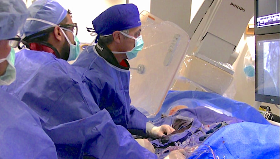
Market Trends in Interventional Angiography Systems

Within the interventional X-ray market, interventional cardiology (IC) was more negatively impacted by COVID-19 than interventional radiology (IR) due to heavier reliance on elective procedures. An increase in the number of structural heart procedures performed and elective percutaneous coronary interventions (PCI) continues to be a factor maintaining clinical demand for the IC market. 

While PCI procedures are assumed to be critical in treating acute myocardial infarctions (MI), only a small amount of PCI procedures performed on patients were for these life-threatening conditions. The rest of the PCI procedures are considered as “elective procedures,” and a large number were deferred during the pandemic. Globally, hospitals saw a decline in cardiovascular procedures. This also includes fewer critical MI PCIs because patients are afraid to go to the hospital and contracting COVID, so many times are ignoring heart attack symptoms.

The IR market has experienced a continued expansion of clinical procedures being performed. An increased incidence of peripheral vascular disease (PAD) is driving demand for general vascular angiography. Despite an estimated 26% drop in the hybrid operating room segment in 2020, fastest growth is predicted for this product category through to 2024.

The pandemic is having a negative impact across all interventional markets (including IC and IR), due to a combination of reduced vendor sales activity (on-site demonstrations, new product launches, conferences) and plummeting elective procedure volumes during national lockdowns.

Angiography imaging system market trends


Key Regional Trends in Interventional Imaging Systems

North America: The North American market has been significantly impacted due to postponement of non-critical surgeries, and as budgets for non-diagnostic X-ray equipment were diverted to COVID response. The North American market will be driven by replacements of existing equipment and increasing demand from outpatient clinics and facilities.

Latin America: Brazil has been one of the worst impacted countries globally by COVID-19, resulting in a severe market retraction for both the interventional and surgical X-ray markets in 2020.

Western Europe: The Western Europe market is forecast to experience pricing pressures from 2020 onwards, with the economy suffering as a result of high COVID-19 impact. Recovery is expected to be steady and gradual through to 2024 as economies struggle with the impact of the pandemic.

Eastern Europe, Middle East and Africa: The Saudi Arabia-Russia oil price war and the COVID-19 pandemic have further setback the prospects of a recovery and the market is forecast to sharply decline in 2020 by 20.1%. Postponement of elective procedures contributed to the huge drop in demand in 2020.

Asia Pacific: The Chinese market has had one of the quickest recoveries from the COVID-19 pandemic globally, with a forecast contraction of high single digits in 2020 for the surgical X-ray market. Demand for hybrid operating rooms and 3-D mobile C-arms is high in China, mainly in high-end hospitals.

Here are some additional market trends:

About the Author: Bhvita Jani is a senior market analyst at Signfy Research. She is part of the medical imaging team focusing on the X-ray market. She brings with her 3 years' experience covering X-ray, MRI and CT research at IHS Markit. She previously worked as an assistant clinical psychologist. She received her bachelor's degree in biology and psychology from Aston University. Signify is a healthcare market research firm based in the U.K.  For more information: www.signifyresearch.net


Viewing all 137 articles
Browse latest View live